
Salivary Gland Stones: Causes, Symptoms, and Treatment Methods
Salivary glands are structures that play a vital role in oral health by producing secretions that aid digestion. However, due to various reasons, hard mineral deposits can form in the salivary ducts, a condition known as salivary gland stones (sialolithiasis). Decreased salivary flow, thickening of saliva, and changes in pH can contribute to stone formation. These stones most commonly occur in the submandibular glands and may cause swelling, pain, and infection over time.
What Causes Salivary Gland Stones?
Although the exact cause of salivary gland stones is not fully known, several factors are believed to contribute:
- Decreased salivary flow rate: Reduced flow can lead to mineral accumulation, facilitating stone formation.
- Increased saliva density: The submandibular glands produce thicker saliva, increasing the risk of stone formation.
- Dietary habits: Low fluid intake and lack of foods that stimulate saliva (like acidic foods such as lemon) can promote stone formation.
- Changes in saliva pH: Reduced acidity can cause minerals to precipitate.
- Certain systemic diseases: Conditions like gout, which cause uric acid accumulation, are associated with higher risk.
What Are the Symptoms?
Symptoms vary depending on the size, location, and severity of the blockage. The most common symptoms include:
- Sudden swelling and pain in the submandibular area during or after eating
- Tenderness under the tongue, in front of the ears, or under the jaw
- Reduced or absent saliva flow
- Recurrent episodes of swelling in the affected area
- Fever and pus discharge if bacterial infection develops
Pain is especially noticeable when eating foods like lemon that stimulate saliva production. If the stone fully blocks the duct, swelling can persist and lead to infection.
How Is It Diagnosed?
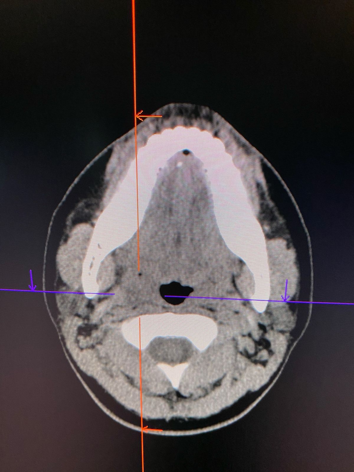
Diagnosis is typically made based on the patient’s symptoms and physical examination, but imaging techniques are used to determine the stone’s size and location:
- Ultrasonography: The most commonly used method, easily identifies the stone and its location.
- CT Scan: Preferred for detecting smaller stones and offers high accuracy.
- Sialography: Involves injecting contrast into the ducts for detailed structural imaging.
- MR Sialography: A radiation-free, advanced imaging method used for assessing salivary gland diseases.
During examination, if the stone is near the duct opening into the mouth, it may be felt manually. If an infection is present, pus may be expressed from the duct upon pressure.
Treatment Options for Salivary Gland Stones
Treatment depends on the size of the stone, degree of blockage, and presence of infection.
Medical Treatment
- If a bacterial infection is present, antibiotics and anti-inflammatory medications are used.
- Increasing fluid intake helps promote natural stone expulsion.
- Foods that stimulate saliva (e.g., lemon, chewing gum) may assist in natural removal.
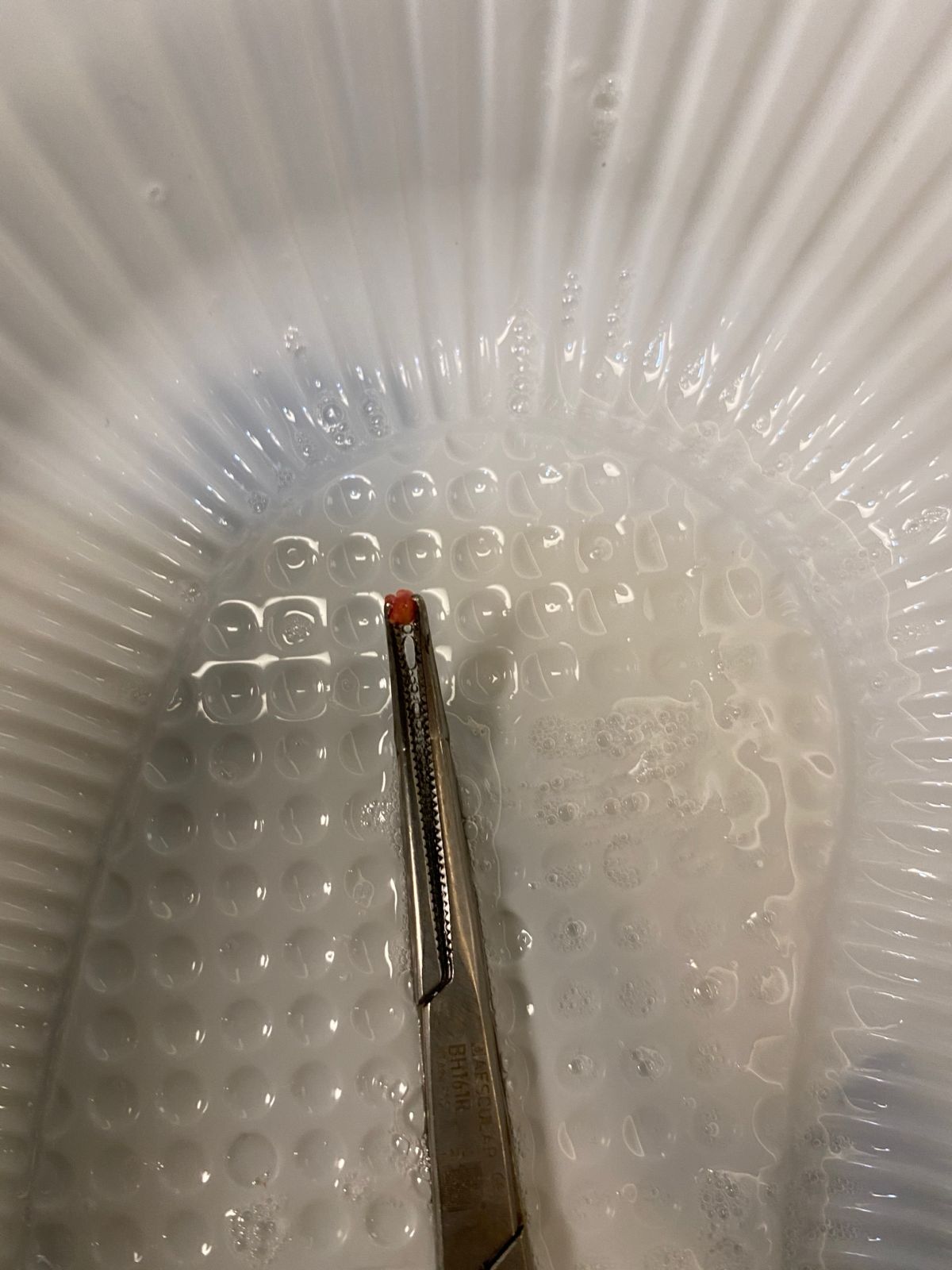
Manual Extraction (Non-Surgical)
If the stone is near the mouth opening of the duct, it can sometimes be removed manually by applying gentle pressure. This method is usually ineffective for larger stones.
Sialendoscopy (Minimally Invasive Technique)
Today, sialendoscopy is a modern, minimally invasive method used to treat salivary gland stones.
What Is Sialendoscopy?
- A thin, flexible camera (endoscope) is used to examine the salivary ducts in detail.
- Small stones can be removed using special tools through the endoscope.
- Larger stones can be fragmented using laser or ultrasonic waves.
Advantages of Sialendoscopy
- Less invasive than open surgery.
- Minimal tissue damage and faster recovery time.
- Can be performed under local anesthesia without general anesthesia.
- Preserves the salivary gland, avoiding unnecessary removal.
Open Surgery
If the stone is large and lodged deep within the duct, surgery may be necessary. In submandibular gland cases, a small incision inside the mouth can be made to remove the stone. In more complex cases, complete gland removal (salivary gland excision) may be required.
How to Prevent Salivary Gland Stones
- Drinking plenty of water helps prevent stone formation by stimulating saliva flow.
- Maintaining good oral hygiene reduces infection risk.
- Consuming foods that stimulate saliva (e.g., lemon, gum, sour fruits) provides natural protection.
- Avoiding smoking and excessive caffeine intake helps prevent saliva thickening and reduces the risk of stones.
Conclusion
Salivary gland stones are generally manageable with early diagnosis and appropriate treatment. Small stones may pass naturally or with minor interventions, while larger stones may require sialendoscopy or surgery. If you experience pain and swelling under the jaw during meals, consult an ENT specialist for early diagnosis and treatment.
