Video-Fluoroscopic Swallowing Study (VFSS): Definition, Procedure, and Clinical Importance
The Video-Fluoroscopic Swallowing Study (VFSS) is a dynamic radiological imaging technique used to evaluate swallowing functions and detect swallowing disorders. This test analyzes the oral, pharyngeal, and esophageal phases in detail, helping to determine how food travels from the mouth to the stomach and whether the airway is adequately protected. VFSS is considered one of the gold standard tests for diagnosing and managing dysphagia (swallowing disorders).
Who Is VFSS Performed On?
VFSS is used to assess whether individuals suspected of having swallowing disorders can safely swallow food and liquids. It is frequently used in the following cases:
- Neurological Diseases: Conditions such as stroke, Parkinson’s disease, ALS, MS, and cerebral palsy can cause dysphagia due to the loss of function in oropharyngeal muscles.
- Head and Neck Cancers: Post-surgical or radiotherapy-related weakening of oropharyngeal muscles and impairment of swallowing reflex are common issues.
- Traumatic Brain Injury and Head Trauma: Damage to the brainstem or cortical centers can weaken or abolish the swallowing reflex.
- Gastroesophageal Reflux Disease (GERD) and Esophageal Strictures: Used to evaluate the functional and structural condition of the esophagus.
- Swallowing Function Assessment in the Elderly: In individuals at risk of dysphagia due to age-related muscle weakness (presbyphagia), VFSS helps determine the safest food consistency.
How Is VFSS Performed?
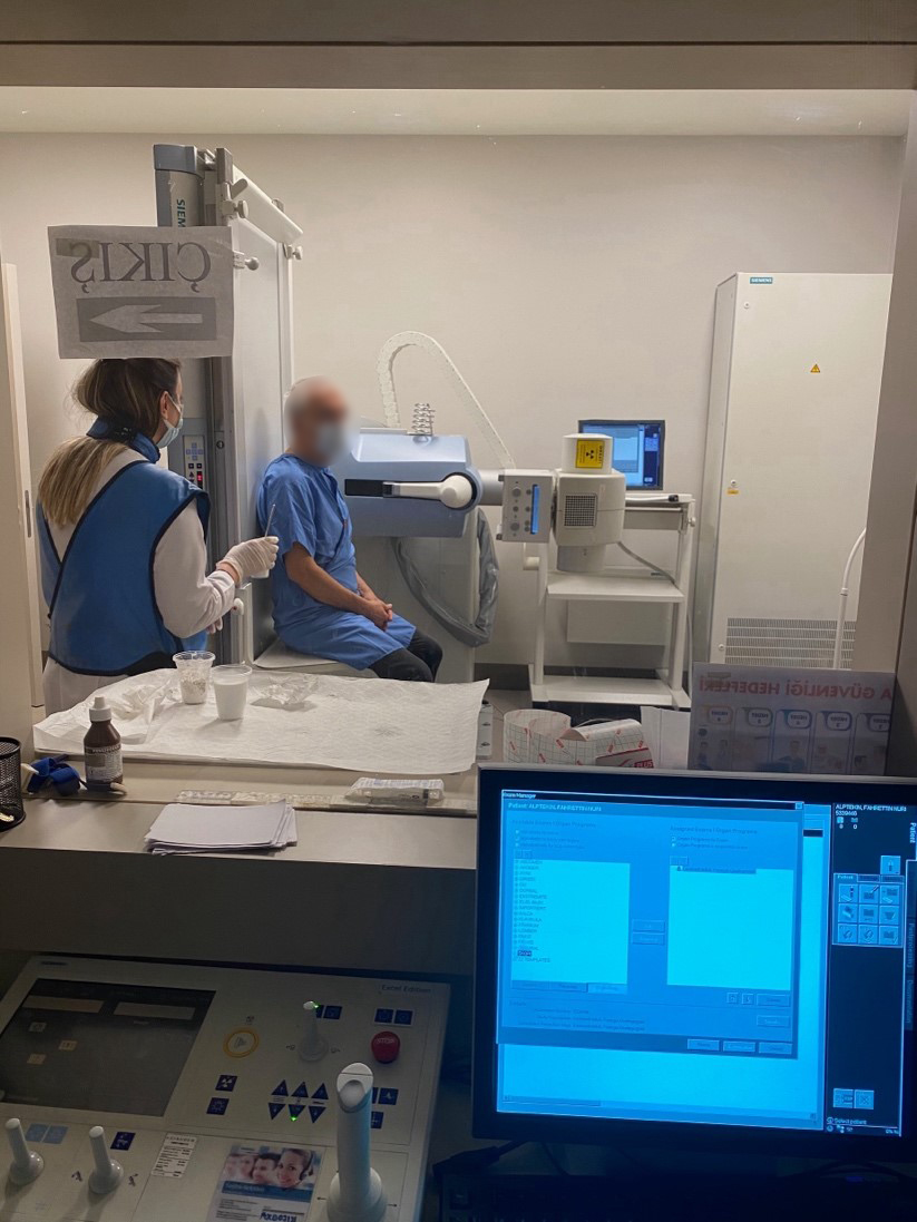
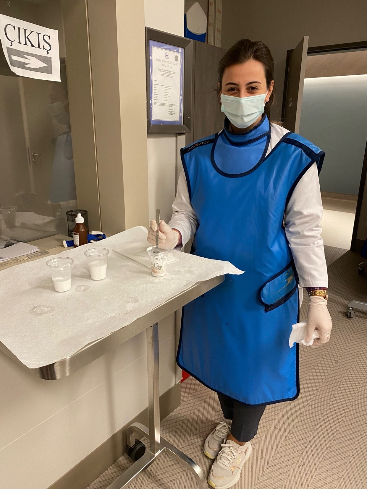
VFSS is a procedure performed collaboratively by a radiologist, a speech-language-swallowing disorders specialist, and an ENT specialist. The procedure includes the following steps:
Patient Preparation and Positioning
- The patient is seated in front of the fluoroscopy device.
- Since the patient will be required to consume foods and liquids of different consistencies, fasting for 4–6 hours may be advised beforehand.
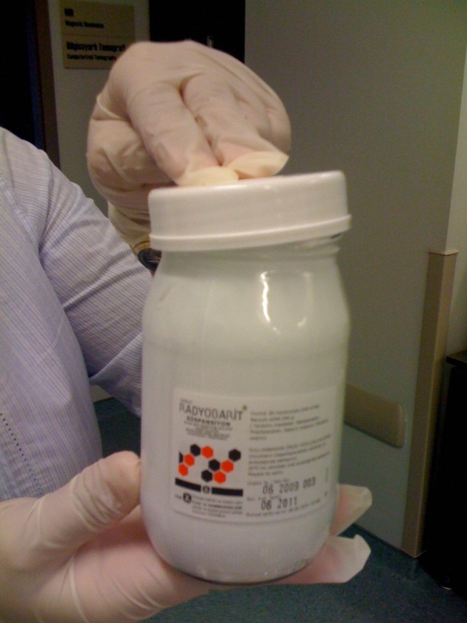
- The patient is given contrast-containing foods (barium suspension) during the radiological imaging.
Dynamic Imaging of the Swallowing Process
- The fluoroscopy device records the swallowing process in real-time from the mouth to the stomach.
- The patient consumes foods in various consistencies and volumes (e.g., water, purée, jelly-like foods, solids).
- In the oral phase, the handling of food by the tongue and its transport to the pharynx are evaluated.
- In the pharyngeal phase, epiglottic closure, laryngeal elevation, velopharyngeal closure, and aspiration risk are assessed.
- In the esophageal phase, how the food passes into the stomach and whether any esophageal dysfunction exists is examined.
Evaluation of Swallowing Disorders and Aspiration Risk
- Whether food enters the airway (aspiration) or pharyngeal residue remains is analyzed.
- Delayed swallowing reflex, epiglottic closure problems, or insufficient laryngeal elevation are identified.
- The safest food consistency for the patient is determined, and an individualized feeding plan is created.
Patient Recommendations and Rehabilitation Plan
- The speech therapist recommends appropriate swallowing maneuvers and exercises to improve swallowing functions.
- The safest way of eating for the patient is determined and dietary modifications are made.
- If necessary, postural adjustments or neuromuscular stimulation techniques may be applied.
Advantages and Clinical Importance of VFSS
VFSS allows objective evaluation of swallowing disorders and is a critical diagnostic tool for increasing patient feeding safety. Its advantages include:
- Evaluates all phases of swallowing dynamically: Oral, pharyngeal, and esophageal phases can be analyzed simultaneously.
- Real-time aspiration monitoring: Determines if silent aspiration is present.
- Effectiveness of compensatory swallowing techniques is tested: Observes if maneuvers reduce aspiration risk.
- Helps create an individualized feeding plan: Determines the safest consistency and feeding method, reducing the risk of aspiration pneumonia.
- Can be combined with other diagnostic methods: Can be used alongside laryngoscopic or manometric assessments.
Differences Between VFSS and FEES (Fiberoptic Endoscopic Evaluation of Swallowing)
| Feature | VFSS (Video-Fluoroscopic Swallowing Study) | FEES (Fiberoptic Endoscopic Evaluation of Swallowing) |
| Imaging Technique | Uses radiological fluoroscopy. | Uses flexible nasal endoscopy. |
| Swallowing Phases Evaluated | Oral, pharyngeal, and esophageal phases can be observed. | Only oral and pharyngeal phases are observed. |
| Direct Aspiration Visualization | Yes, airway penetration can be visualized. | Evaluated indirectly (e.g., with blue dye test). |
| Radiation Exposure | Yes, but at minimal levels. | No radiation exposure. |
| Bedside Applicability | No, performed in a radiology unit. | Yes, can be done in outpatient clinics or at bedside. |
Conclusion and Clinical Value
Video-Fluoroscopic Swallowing Study (VFSS) is one of the most comprehensive methods for analyzing swallowing disorders. This technique is essential for identifying safe feeding strategies, reducing aspiration risk, and individualizing swallowing therapy. Early diagnosis and proper treatment planning in individuals with dysphagia are critical for preventing serious complications such as nutritional deficiencies and aspiration pneumonia.
Barium
Barium-Enhanced Foods of Various Consistencies (Prepared for Testing)
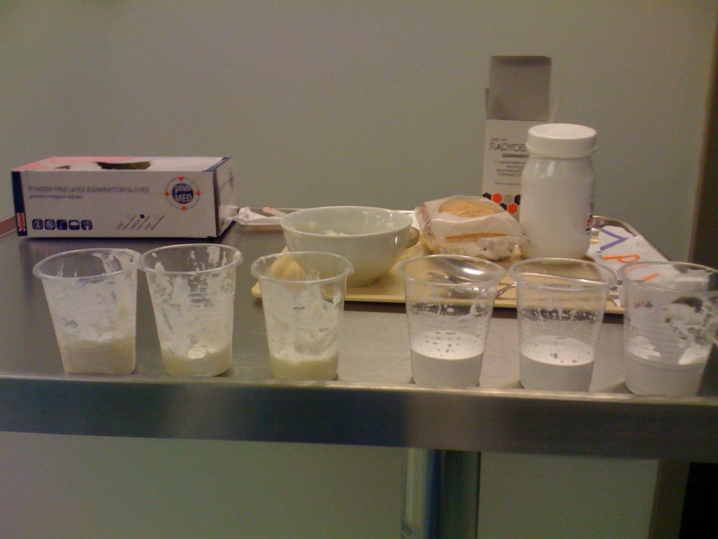
Barium-Enhanced Pudding

Speech-Language-Swallowing Specialist During VFSS
VFSS